

The 20-week ultrasound: A guide to your pregnancy anatomy scan
Get ready for your detailed pregnancy ultrasound. We explain what the 20-week anatomy scan checks for, how long it takes, what the results mean, and why a follow-up is common.

If your pregnancy is approaching your 20 weeks, you've probably heard about the anatomy scan. Or your doctor/midwife has recommended you for one. First of all, there’s nothing to be anxious about, it’s a routine check for your growing baby. While this is optional, it’s important to do the scan to understand any possible challenge.
Don’t focus on negative stories where things go wrong, only about 3% of abnormalities occur. So relax, and arm yourself with the necessary info.
Let’s walk through what this scan is, what happens during it, and what you can expect.This scan is one of the most detailed check-ups during pregnancy. Here’s a simple, honest guide to help you know exactly what to expect.
{{button}}
The 20 week Anatomy Scan
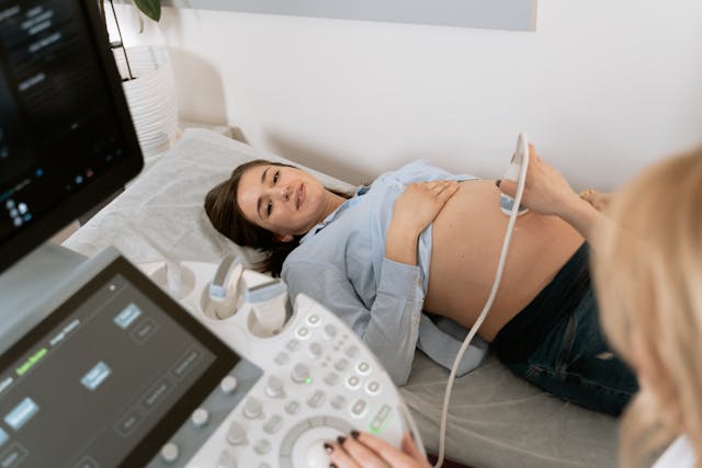
The 20 week anatomy scan is a comprehensive ultrasound to check for your baby’s development, your uterus, placenta and the amniotic fluid. It’s an in-depth examination of your baby's development. It’s also called the second-trimester anatomy scan, 20-week ultrasound or anomaly scan. Unlike your earlier ultrasounds, this one takes a comprehensive look at your baby's organs, bones, and overall structure to make sure everything is developing as it should. You’ll also get to know you baby’s gender if you wish.
The scan focuses on detecting fetal structural defects and markers for chromosomal or genetic abnormalities. Currently, about 50% of all fetal abnormalities are detected prenatally, so this scan is your best opportunity for early detection.
When Is the Anatomy Scan Done?
The anatomy scan is typically scheduled between 18 and 23 weeks of pregnancy. The ideal time is right around 20 weeks, when your baby is developed enough to see the structures clearly, but there's still time to make informed decisions if any issues are found.
How Long Does the Anatomy Scan Take?
The anatomy scan takes between 30 minutes to an hour. The length really depends on a few factors: your ultrasound technician's pace, how cooperative your baby is, and your own comfort. If you need to shift positions frequently, the scan will naturally take a bit longer. It's really in-depth, so don't be surprised if it feels thorough.
What Does an Anatomy Scan Cover?
During your appointment, the sonographer will take pictures and measurements of many parts of your baby's body such as:
- Heart (including blood flow and all four chambers)
- Brain, head, neck, and spine
- Kidneys and bladder
- Arms and legs
- Hands, fingers, feet, and toes
- Lips, chin, nose, eyes, and face
- Chest and lungs
- Stomach and intestines
They’ll also check for:
- Fetal heart rate
- Umbilical cord blood flow and its attachment to the placenta
- Position of your placenta
- Amount of amniotic fluid
- Your uterus, ovaries, and cervix
- The cord to confirm the appropriate number of arteries and vessels,
- Measure your baby's bones.
Conditions Screened for
There are 11 essential conditions that are checked during the 20 week scan. These conditions include:
- Anencephaly
- Open spina bifida
- Cleft lip and palate
- Diaphragmatic hernia
- Gastroschisis
- Exomphalos
- Serious cardiac abnormalities
- Bilateral renal agenesis
- Severe skeletal dysplasia
- Edwards' syndrome (trisomy 18)
- Patau's syndrome (trisomy 13)
Some conditions can be seen more clearly than others, so the scan may not find everything. But screening is the best way to identify any potential issues early, helping you and your doctors make the best decisions for you and your baby.
Understanding Detection Rates
The anatomy scan has a high detection rate for major structural malformations, ranging from 15% to over 90% depending on what's being examined. Detection rates vary by organ system, and factors like equipment quality and sonographer experience play a role in accuracy.
What the evidence shows:
- The scan may improve detection of multiple pregnancies
- It may increase detection of major fetal abnormalities before 24 weeks (about 3.45 times more likely to detect compared to no routine scan)
- It may reduce induction of labor for post-maturity, likely because it provides more accurate dating
- Studies show the scan probably makes little difference to perinatal loss rates
- Long-term follow-up shows no harm to children's physical or intellectual development from ultrasound exposure
Fetal structural anomalies affect up to 3% of pregnancies. While the scan isn't 100% effective, it's safe and valuable. It empowers you to make informed decisions and can improve some pregnancy outcomes by enabling early detection, planning for delivery at the right time and place, and giving you time for emotional preparation.
What to Expect at Anatomy Scan
Here's something to bear in mind. The sonographer won't give you detailed comments, especially if they see something unusual. For example, they might point out the heart, legs, or head, but they won't give you details about whether everything is normal or abnormal during the scan itself.
This is standard practice so don’t worry. The findings will be written in a report for your obstetrician, who will explain your results at a follow-up appointment. If something needs further investigation, you may be referred to a specialist at a fetal medicine department to confirm or rule out the sonographer's findings.
Note: Good technicians often comment on and compliment positive findings, which can be comforting during the scan.
The scan is done completely over your belly, however, there might also be a transvaginal to measure your cervix.
About Your Placenta Position
During the scan, they'll tell you where your placenta is positioned:
- Anterior placenta: The placenta is at the front of your uterus. You might not feel as much movement early on because the placenta cushions you from baby's kicks.
- Posterior placenta: The placenta is at the back. You may feel more movement.
Neither position is better or worse, they're just different and have no medical significance.
The one exception is a low-lying placenta (close to your cervix). If this is found, don't panic. In the vast majority of cases, the placenta moves up as your uterus grows. Your doctor will check again later in pregnancy. Only if it remains very close to your cervix would you potentially need a cesarean section.
When Baby Doesn't Cooperate
It’s highly common that the sonographer won't get all the measurements they need, and you'll have to come back a week or two later. This happens all the time and doesn't mean anything is wrong.
If your baby is in an awkward position, moving too much, or just being uncooperative, they simply can't get a good look at everything they need to see. So you’ll need a follow-up scan.
The Bottom Line
The 20-week anatomy scan is a milestone in your pregnancy journey. It gives you and your healthcare team valuable information about your baby's development and helps ensure the best possible care. While waiting for results can feel stressful, remember that this scan is designed to give you answers and options.
Technological advancements continue to improve what we can see and detect, making this scan an essential part of prenatal care. It's not perfect, but it's one of the best tools we have to monitor your baby's health and prepare for their arrival.
{{pink-banner}}


.avif)
