

13 week ultrasound: The procedure and results to expect
The 13-week ultrasound combines nuchal translucency screening with anatomy basics. Here's how the scan works, what it can detect, and what results mean.

13 weeks is typically considered the final week of the first trimester. By this time, your baby is already about the size of a peach (7.4 cm long) and continues to develop rapidly. As you transition to the second trimester, you might notice an improvement in your early pregnancy symptoms, feel more energized, and expect to have your first-trimester screening soon.
A 13 week ultrasound can be your first routine screening. However, in some cases, you may have had the first scan earlier, and repeat the procedure at 13 weeks to confirm and detail previous findings. At this time, the doctor will assess:
- The baby’s location
- Gestational age
- Due date
- The number of fetuses
- Growth and development
The exciting news is that your baby is already grown and has more detectable features at 13 weeks. That is, you can expect to see its body, head, some facial features, movement, and heartbeat in an ultrasound.
{{button}}
13 weeks is the final week of your first trimester. This is an incredibly exciting, yet stressful, transition period that’s associated with many important milestones. At this time, the unpleasant early pregnancy symptoms, such as morning sickness, should start improving or even go away completely. Likely, you will also feel more energized. And for sure, you will feel very excited about what comes next.
One of the most important stages during this period is a 13 week ultrasound. If you didn’t have your routine or indicated screening earlier, this will be the first time you will see your baby and learn more details about your pregnancy development. So, what should you prepare for? Let’s figure it out!
Why Do You Need to Have a 13 Week Ultrasound?
You can experience a 13 week ultrasound both as the first pregnancy screening and as a repeated one.
The truth is that there is no universal practice related to the timing and frequency of pregnancy ultrasound. The earliest one can be done at 5 weeks, and it’s often performed in high-risk pregnancies and if there are any medical indications, such as bleeding or pain. Also, it’s common to have your first screening between 6-9 weeks as part of routine prenatal care. In this case, a 13 week ultrasound can act as a repeated screening. It can be used to confirm gestational age and follow up on previous scan findings.
In other cases, it’s possible that your 13 week ultrasound is your first-ever scan. Some doctors prefer to wait until a baby is more developed to be able to perform a more accurate assessment. This is often done in low-risk pregnancies, when there are no reasons for early appointments. In this case, your doctor will use this scan to detect the following:
- The baby’s location
- Gestational age
- Due date
- The number of fetuses
- Growth and development
Additionally, your healthcare provider will examine the fetal structure to rule out any anomalies. They will check bones, limbs, and major organs.
The Nuhal Scan
During a 13 week ultrasound (especially if it’s your first screening), you will also likely undergo the Nuchal Translucency (NT) scan. This is one of the most essential screenings in early pregnancy that assesses the size of the fluid-filled space at the back of your baby’s neck. It can be conducted anywhere between 11 weeks and 13 weeks.
The NT measurement allows doctors to identify the possibility of a chromosomal condition, such as Down syndrome, Edwards syndrome, and Patau syndrome.
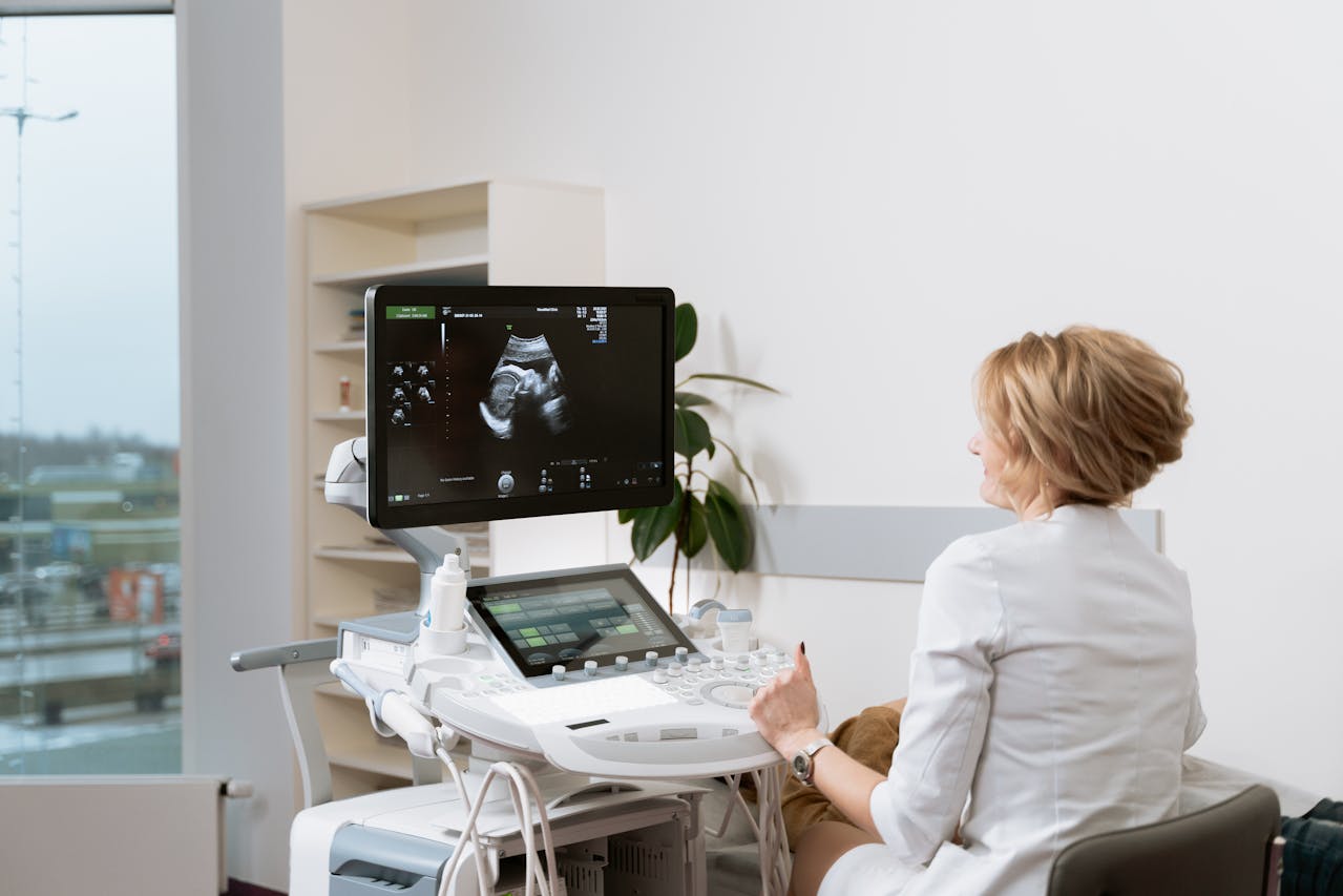
Understanding a 13 Week Ultrasound Procedure
Now, it’s clear that having an ultrasound 13 weeks pregnant is an important part of normal prenatal care. So, what can you expect from it?
As you may know, there are two types of ultrasound used in pregnancy – transvaginal and abdominal. Traditionally, doctors prefer to do transvaginal probes during early scans, such as at 7 weeks. However, since at 13 weeks your baby is already somewhat grown, this procedure is typically performed using an abdominal ultrasound.
The process goes like this:
- You will be taken to the ultrasound room.
- The sonographer will apply gel to your abdomen.
- The specialist will move the transducer around your belly to get an image.
On average, this procedure takes around 30 minutes.
Important note: Although it’s more common to use an abdominal ultrasound, a doctor may also use a transvaginal one. This usually happens when they need a more detailed image of the fetus and your internal organs for a more detailed examination. Also, it’s common to use a transvaginal ultrasound in women with increased BMI if the sonographer fails to receive a clear image. This is a normal practice, so don’t get worried if you are suggested this kind of scan.
What Does a 13 Week Ultrasound Look Like?
So, now you know why screening at 13 weeks is important and how it is performed. Finally, we’ve gotten to the most exciting part. How can you expect to see your baby at 13 weeks ultrasound?
The good news is that now your baby looks more like a baby. The size of the fetus should be around 7.4 cm. This is close to the size of a peach. It already has a formed body and head, and even some facial features. So, there is quite a lot that you can see.
Typically, a healthy 13 week ultrasound should show you the following:
- Head and face – The baby still has a rather large head, but it already has some noticeable facial features, such as a nose, eyes, and a mouth.
- Body and limbs – As you reach 13 weeks, your baby already has a distinguished shape with a clearly visible body and long, thin limbs. If the image is detailed, you might be able to see tiny fingers and toes. The spine and ribs are also visible.
- Movement – You may not feel it yet, but your baby is getting very active around 13 weeks. So, when you are looking at a sonogram, it’s common to see different movements, as your baby stretches or moves its limbs.
On top of seeing your baby, you should also see or hear their heartbeat while doing an ultrasound 13 weeks pregnant. A doctor can assess it with an ultrasound or by using a Doppler. The normal heart rate at this time ranges between 140 and 170 beats per minute (bpm).
Detecting the Gender of Your Baby at 13 Weeks Ultrasound
As you’re moving closer to your second trimester, you are probably already looking forward to learning whether you are having a boy or a girl. This is an important and emotional moment for the majority of parents-to-be. So, it’s not odd if you’re feeling a strong urge to learn the sex as soon as possible.
The question is: Can you actually do it during a 13 week ultrasound? The good news is that it’s generally possible. 11 weeks is the earliest time when a healthcare provider can assess your baby’s gender. Typically, the doctor will use the “nub theory” for this purpose. This theory uses the angle of the genital tubercle (nub) to determine the gender. When it’s angled upward, you might have a boy, and when it has a parallel or downward angle, it suggests a girl. Yet, it’s important to remember that your baby is still quite small and is developing at 11 weeks, which means that the accuracy of such evaluation is still low. The good news is that it increases along with your gestational age, and 13 weeks is the time when it becomes quite accurate.
According to studies, a 13 week ultrasound enabled doctors to predict male fetuses with 84% accuracy, and female fetuses with 71% accuracy. So, yes, there is a chance that you can learn your baby’s sex early. However, it’s important to set realistic expectations. Since the accuracy is not 100%, it can’t be called a definitive diagnosis yet.
FAQ
Is 13 weeks a common time for an ultrasound?
Yes, 13 weeks is considered a good time for a routine pregnancy ultrasound. At this time, you are in the final week of your first trimester. Your baby is already grown and well-developed. So, many doctors use this time for the first-trimester screening, during which they confirm your gestational age, identify the number of fetuses, and assess the potential risks. However, it’s also possible that it’s not the first, but a repeated scan, especially if there were any medical indications for an early ultrasound.
Why can doctors use a transvaginal approach during a 13 week ultrasound?
Most often, you should expect an abdominal ultrasound 13 weeks pregnant. However, sometimes, a healthcare provider may recommend a transvaginal scan instead, and it’s okay. This approach can be taken if there is a need for a closer look at your pelvic organs and fetus. It can be important if the doctor needs to check for any maternal anatomical abnormalities or measure the length of the cervix. Also, a transvaginal ultrasound might be preferred in women with a higher BMI or obesity because an abdominal ultrasound may not provide a clear image in this case.
Can the doctor detect the baby’s gender at 13 weeks?
Yes. The earliest time when healthcare providers can determine the baby’s gender is 11 weeks. Doctors use the “nub theory” to make an informed prediction, and its accuracy increases along with your gestational age. There is a chance that your healthcare provider will be able to accurately detect your baby’s sex during a 13 week ultrasound. However, you may want to wait longer to get the most accurate result.
Conclusion
A 13 week ultrasound is considered a good time to have your first-trimester scan. If you haven’t had an ultrasound before, your doctor will use it to locate the baby and calculate your due date. Additionally, they will likely assess the fetus’s structure and perform the Nuhal scan to rule out any chromosomal conditions.
After reading this guide, you should have a better understanding of this procedure, its importance, and the results you can expect to see. Use this knowledge to prepare for this emotional moment and reduce the anxiety.
{{pink-banner}}

.jpg)


.avif)
